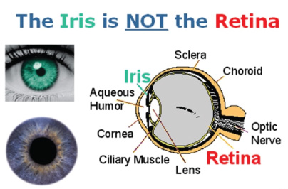
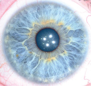
The iris (plural: irides or irises) is a thin, circular structure in the eye, responsible for controlling the diameter and size of the pupils and thus the amount of light reaching the retina. “Eye color” is the color of the iris, which can be green, blue, or brown. In some cases it can be hazel (a combination of light brown, green and gold), grey, violet, or even pink. In response to the amount of light entering the eye, muscles attached to the iris expand or contract the aperture at the center of the iris, known as the pupil. The larger the pupil, the more light can enter. Iris recognition is an automated method of biometric identification that uses mathematical pattern-recognition techniques on video images of the irides of an individual’s eyes, whose complex random patterns are unique and can be seen from some distance.
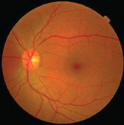
Unlike retina scanning, and iris scan uses camera technology with subtle infrared illumination to acquire images of the detail-rich, intricate structures of the iris. Digital templates encoded from these iris scan patterns by mathematical and statistical algorithms allow unambiguous positive identification of an individual. Databases of enrolled templates are searched by matcher engines at speeds measured in the millions of templates per second per (single-core) CPU, and with infinitesimally small False Match rates. Hundreds of millions of persons in countries around the world have been enrolled in iris recognition systems, for convenience purposes such as passport-free automated border-crossings, and some national ID systems based on this technology are being deployed. A key advantage of iris recognition, besides its speed of matching and its extreme resistance to False Matches, is the stability of the iris as an internal, protected, yet externally visible organ of the eye.


